What This Document Is
This is a detailed exploration of the anatomy of the pharynx, a crucial component of the human speech mechanism. Specifically designed for students in advanced Speech-Language Pathology courses, this resource delves into the structural organization of this vital passageway. It focuses on identifying and understanding the distinct regions of the pharynx and the musculature responsible for its function. The material is sourced from university-level coursework (University of Illinois at Urbana-Champaign, SHS 300) and builds upon foundational anatomical knowledge.
Why This Document Matters
This resource is invaluable for students preparing for careers in speech-language pathology, audiology, or related fields. A thorough understanding of pharyngeal anatomy is essential for diagnosing and treating a wide range of communication and swallowing disorders. It’s particularly helpful when studying the physiological processes of respiration, phonation, and swallowing. Students will find this material beneficial during coursework, while preparing for exams, or as a reference during clinical practicums. It provides a strong foundation for understanding how structural elements contribute to functional abilities.
Common Limitations or Challenges
This document focuses *solely* on the anatomical structures of the pharynx. It does not provide in-depth coverage of related physiological processes like swallowing mechanics or detailed neurological control. While it touches upon functional implications, it does not offer clinical applications or treatment protocols. Furthermore, it assumes a baseline understanding of general anatomical terminology and directional concepts. It is intended to be a focused anatomical study, not a comprehensive guide to speech or swallowing disorders.
What This Document Provides
* Detailed descriptions of the three primary divisions of the pharynx: nasopharynx, oropharynx, and laryngopharynx.
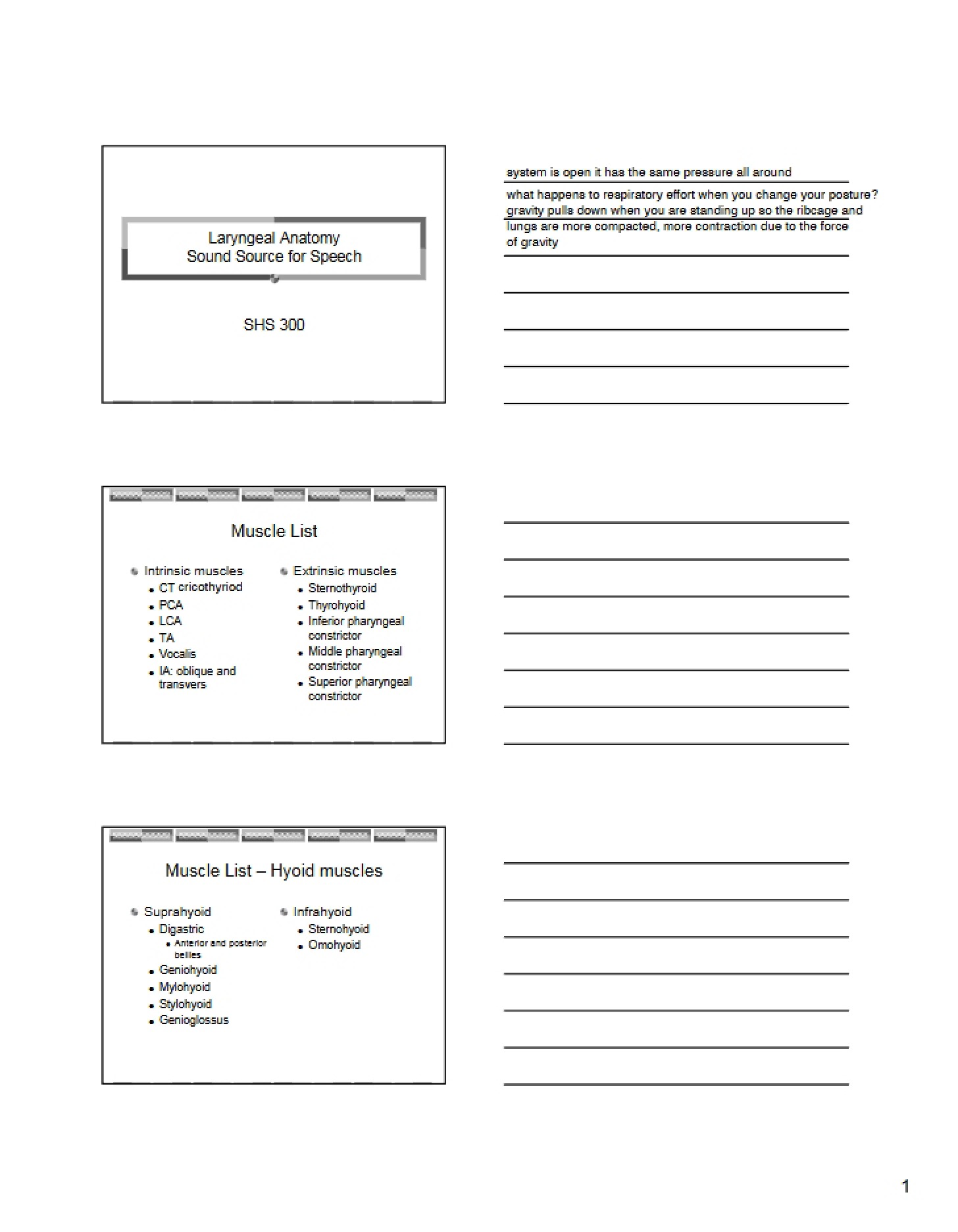
* An overview of the key muscles associated with pharyngeal function, including the constrictor muscles and other supporting structures.
* Identification of important anatomical landmarks and boundaries within each pharyngeal region.
* Discussion of specific anatomical features like the Eustachian tube orifice and various pharyngeal raphes.
* References to external visual resources to aid in spatial understanding of the anatomy.